Wednesday 27 May 2015

Ulota crispa

Slow but steady progress on bryophytes. Not a good time of year to be studying them as most are dried out but there are still one's that can be done. This Ulota crispa was characterised by the curled distorted leaves when dry and the hairy calyptra (sheath over the capsule).

Ulota crispa leaf

Ulota crispa mid leaf cells

Ulota crispa basal leaf cells

Ulota crispa leaf tip

Ulota crispa

Friday 22 May 2015

Eulophidae

Found this tiny 1-2mm long hymentoptera on 20/5/15 in Sand Bay, North Somerset which has been identified as a Eulophidae sp. I tried to narrow it down further and think it may be a Entedon sp.

Eulophidae pos Entedon sp

Wednesday 20 May 2015

Campylopus introflexus

Found in a flower pot in the back garden this species is said to have reflexed hairpoints when dry.

Campylopus introflexus capsule

Campylopus introflexus

Campylopus introflexus leaf

Monday 18 May 2015

Orthotricum anomalum

This specimen is quite dried out but had few new growths so thought it would be worth a go. After some three hours of deliberation I came up with the conclusion that it is Orthotrichum anomalum

Orthotrichum anomalum peristome teeth

Orthotrichum anomalum

Orthotrichum anomalum leaf

Tuesday 12 May 2015

Sarcophaga carnaria?

Literature is not easy to get hold of for Sarcophagidae. S.carnaria is one of the more common of the group which I believe is photographed here.


Phaeographis dendritica

A surprise to see brown septate spores on this lichen under the microscope. Although a fairly common lichen it is not that easy to find in my county so a good record.

Phaeographis dendritica plus spore in inset

Fuscidea cyathoides

This lichen showed many spores that appear to be kidney shaped 11µm a characteristic of Fuscidea cyathoides. The ascus KI reaction I could see was fuscidea type but only after I knew what shape to look for.

Fuscidea cyathoides

Friday 8 May 2015

Fuscidea kochiana

A straight forward identification for this one. Colourless 10x5µm spores turning brown when mature, swollen and pigmented cap of the paraphyses, fuscidea type ascus all useful characters.

Fuscidea kochiana KI ascus reaction

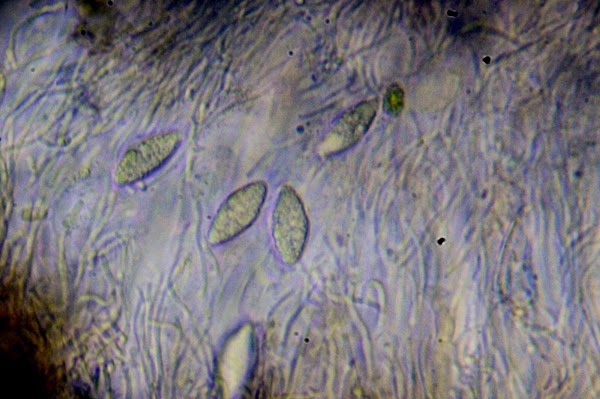
Fuscidea kochiana mature spores.

Fuscidea kochiana

Fuscidea kochiana

Thursday 7 May 2015

unidentified

Found on siliceous rock. Spores 22.5x9µm


Hypogymnia tubulosa

Slightly puzzled by this one due to the extensive isidia. That said I can't see that it can be anything else other than Hypogymnia tubulosa.

Hypogymnia tubulosa

Hypogymnia tubulosa

Graphis scripta

Interesting to compare the spore septum shape of Graphis after having so many Opegrapha's under the microscope lately. The 12-septate spore eliminates Graphis alboscripta and the K- and lack of furrowed apothecia eliminates Graphis elegans.

Graphis scripta spore

Graphis scripta

Acarospora fuscata

A distinctive lichen in the field and under the microscope too. The asci contain 200 or so spores. Narrowing down to species level however was more difficult. This one was on siliceous rock (carboniferous granite). I went for A.impressula because of the C- reaction but the areole size and lack of records for the site forced me to go with A.fuscata in the end.

Acarospora fuscata ascus

Acarospora fuscata

Acarospora fuscata

Wednesday 6 May 2015

Micarea lignaria

Back on the lichen again. This one I thought would be easy with as the spores were 7-septate but despite this it wasn't easy. For me the biggest problem was working out the apothecia type. Lecanorine was easy to eliminate leaving perithecia and lecideine. The lack of a flask left me with lecideine as the only option. The thallus I also had trouble with. The book seem to have trouble describing it also.

Micarea lignaria

Micarea lignaria section

Micarea lignaria spore

Micarea lignaria

Sunday 3 May 2015

Unidentified

One taken from Sand Point back in February. Lack of familiarity with KI ascus types through the microscope is preventing me from identifying this one. I am publishing this one with a view to returning at a later date for a conclusion.
Hymenium: 50µm
Spores: 12x6µm
Thallus: C-, K-

apothecia section (.5mm diamter)

through the microscope

spores

ascus KI reaction

Friday 1 May 2015

Unidentified - Lecanora polytropa?

I thought this one would be easy but no candidates so far. The ascus was KI+ but couldn't identify the reaction so may come back to this one at a later date. When dried out the colour looks different so I suspect that lack of any visual matches has put me off making a confident ID.
Hymenium: 100
Epithecium: 7.5
Spores: 10x7.5